
Perforator flaps are a reconstructive technique that moves a patient’s own skin and subcutaneous fat to cover a wound, defect, or area of missing tissue. Unlike older flap methods, they’re designed to spare the underlying muscle and major vessels, which reduces donor site morbidity the complications that occur at the site where tissue is harvested. The flap stays alive because perforators, small specialised blood vessels that pass through muscle or fascial septa from deeper tissue, continue to supply the transferred skin throughout and after the procedure.
According to Dr. Leena Jain, best plastic surgeon in Borivali, Perforator flaps give us a way to bring healthy, vascularised tissue into wounds where the local bed has already failed and that’s often what turns a non-healing case into a successful reconstruction.
What Makes Perforator Flaps Different from Skin Grafts or Older Flap Techniques?
Skin grafts depend on the wound bed to supply them with blood, which fails when that bed is already compromised, and that’s precisely the gap perforator flaps fill.
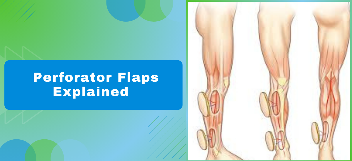
- Blood supply: The flap is raised around a perforating vessel that threads up through muscle to feed the overlying skin, so transferred tissue arrives with circulation already working rather than waiting on the wound bed to nourish it from below.
- Muscle sparing: Older myocutaneous flaps took skin and the full underlying muscle together, but perforator technique dissects only the vessel and the skin paddle, leaving muscle intact and cutting post-operative pain considerably at the donor site.
- Contouring ability: Because the flap can be thinned and shaped before inset, it fits the exact geometry of a defect without adding excess bulk something that matters a lot on the foot, hand, or face where stiffness or volume causes real functional problems.
- No foreign material: The tissue is yours, so immune rejection isn’t a factor, and the body integrates the flap far more predictably than it does with synthetic mesh or cadaveric grafts placed over a compromised bed.
Chest wall defects after cancer resection are one area where perforator-based reconstruction consistently changes outcomes a breast surgery review can confirm whether this approach fits your defect.
How Does the Surgeon Actually Plan and Perform a Perforator Flap?
Planning starts before the patient reaches theatre, and the preoperative mapping stage is honestly where most of the outcome is decided.
- Vessel mapping: A CT angiogram or handheld Doppler locates the perforating vessels at the donor site before surgery, confirming which ones are wide enough and well-positioned to reliably support the planned skin paddle without putting surrounding tissue at risk.
- Flap design: The skin paddle is marked directly over the mapped perforator with a calculated safety margin, and for any plastic surgeon in Bandra handling complex lower limb wounds, the anterolateral thigh stays the most frequently harvested donor site because of its reliable anatomy and low morbidity profile.
- Microsurgical join: In free flap cases the pedicle vessels are divided and reconnected to recipient vessels near the wound under an operating microscope, using sutures finer than a human hair to join vessels that are sometimes under two millimetres wide. It’s painstaking work.
- Post-op checks: Flap perfusion is assessed every hour for the first 48 hours after surgery because venous congestion or early arterial compromise, caught quickly, can usually be reversed before the flap is lost entirely.
The best plastic surgeon in Bandra blog covers more on how wound complexity affects surgical decision-making in reconstructive cases.
Why Choose The Plastic Surgery Clinic?
Dr. Leena Jain brings 7+ years in plastic and reconstructive microsurgery, with direct experience in perforator flap design, free tissue transfer, diabetic foot salvage, and post-trauma limb reconstruction including cases referred after failure at other centres. She trained under internationally recognised microsurgeons, and that technical foundation is visible in how she approaches high-stakes, complex reconstructions.
What patients consistently mention is that they knew exactly what was happening before they went into theatre. Not a vague overview the specific flap chosen, the donor site rationale, the monitoring protocol post-op. That level of detail is uncommon, and it makes a real difference to how patients move through recovery.
FAQs
What is a perforator flap in simple terms?
Skin and fat moved from one body area to repair a wound, kept alive by its own blood vessel throughout.
How long does perforator flap surgery take?
Between four and eight hours depending on flap complexity and whether microsurgical vessel connection is needed.
Is the donor site left with a large scar?
A linear scar remains but muscle is preserved, so long-term donor site problems are far less than older techniques caused.
When can a patient move or walk after this surgery?
Gentle mobilisation typically starts within two to three days, with full activity depending on wound location.
References –
- Perforator Flap Anatomy and Classification — National Center for Biotechnology Information
2. Free Flap Microsurgery Outcomes — PubMed, National Library of Medicine
